Improving Detection of the Lamina Cribrosa
The image quality of commercial optical coherence tomography (OCT) devices has drastically improved and is now sufficient to visualize deep connective tissue structures such as the lamina cribrosa (LC) and the posterior sclera – both of which are thought to be important biomechanical players in ocular disorders such as glaucoma, myopia, and papilledema. However, OCT image quality is still inferior to that of histology, which is a major limitation for clinical hypothesis testing using OCT. Specifically, in most subjects, the LC is only partially detectable even with recent hardware improvement techniques such as enhanced depth imaging (EDI). The partial visibility of the LC is due to light attenuation. This limits the visualization of the anterior, and especially the posterior, boundaries of the LC and the detection of the LC insertion into the peripapillary sclera. Moreover, when light attenuation is too strong, shadows, usually casted by blood vessels from the central retinal trunk, are created which further limit proper LC detection.
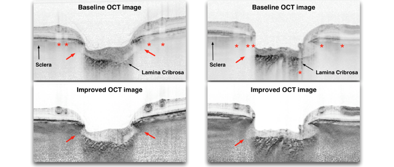
To improve detection of the LC in OCT images, one needs to incorporate the physics of light attenuation into standard OCT theory in order to: 1) evaluate the effects of light attenuation and 2) correct such effects. By adapting and improving compensation techniques that were originally developed for Ultrasound imaging we were able to drastically enhance OCT images of ONH. Specifically, these techniques can 1) improve the visibility of LC/sclera insertion sites (Left Figure; red arrows); 2) improve the visibility of focal LC defects (Right Figure; red arrows); 3) drastically improve contrast across all tissue boundaries (especially the anterior LC boundary; Figure); 4) remove blood vessel shadows in the ONH tissues (Right and Left Figures; red asterisks); 5) help identify the full thickness of the LC (data not shown); and 6) improve the visibility of the peripapillary choroid and sclera (Right and Left Figures). These compensation techniques, when combined with EDI, provide the best LC detection.
In summary, we have demonstrated that OCT image quality can be improved using relatively simple image processing that can correct for light attenuation. We are currently refining these techniques in order to limit noise over-amplification and to provide a better integration with OCT hardware. It should be emphasized that not all of the above improvements can be achieved in every eye, and care must be taken for data interpretation. Although only one SD-OCT (Spectralis, Heidelberg Engineering, Heidelberg, Germany) has been tested here, the proposed techniques can also be applied to time-domain, swept-source and adaptive optics OCT.
This is a project in Collaboration with Dr Jean Martial Mari from INSERM. Note that the original OCT Scans were obtained from Sean Park from the New York Eye and Ear Infirmary.
Related Work
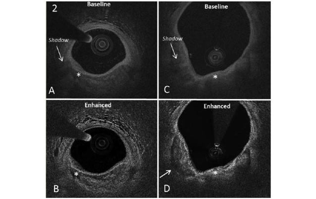
Improving Detection of Coronary Artery Plaques
Compensation techniques are used to improve the visibility of coronary artery plaques.